
Echocardiography for Pets in Punta Gorda FL
Echocardiography, also known as cardiac ultrasound, is one of the most useful diagnostic techniques in veterinary cardiology. Sound waves are directed into the body using a probe. These sound waves then interact with the tissues in the body. Some of the sound waves are reflected back to the transducer. By analyzing these reflected sound waves, the ultrasound machine is able to create images of the heart that are then displayed on the monitor. This allows non-invasive visualization of the heart muscle, the heart valves, and the great arteries. In addition to producing images of the heart, echocardiography can also tell us about heart function by using these various features:
2-Dimensional Echocardiography
This mode produces black and white images of the heart and great vessels. These images can be still frames, or moving images, which show near real-time cardiac motion. 2-Dimensional Echocardiography is used to get an overall impression of cardiac size. Measurements of specific cardiac chambers can be made using the still frame images. This mode also allows rapid detection of free fluid in the chest cavity (pleural effusion) or surrounding the heart (pericardial effusion).
M-Mode Echocardiography
M-Mode is the oldest form of cardiac ultrasound. M-Mode provides a cross-sectional view of the heart shown over time. This allows precise measurement of cardiac chambers through the different phases of the cardiac cycle. Modern echocardiography machines allow for a combination of 2-Dimensional and M-Modes using a split screen display. This helps guide optimal placement of the M-Mode cursor, thereby obtaining the most accurate measurements.
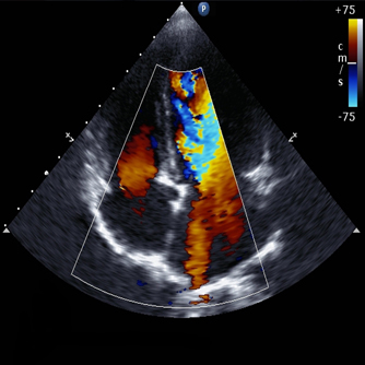
Color Flow Doppler
The color flow feature provides a visual display of blood flow within the heart, using Doppler-based technology to map both the direction and speed of blood flow. The colors are superimposed on the 2-dimensional image, giving an easy way to interpret representation of blood flow within the heart. Abnormal blood flow patterns are readily seen using this feature.
Pulsed Wave and Continuous Wave Doppler
These Doppler features allow us to graph the speed and direction of blood flow within the heart. These modes are used with the color flow Doppler to document leaks in heart valves, abnormal flow between chambers though defects in the heart, and high-speed flow secondary to narrowed regions within the heart.